- +34 971 721 339
- rx@doctorrovira.com
- C\ Missió, 4-B, Bajos
Mammography
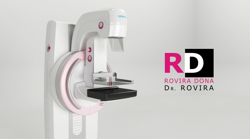
MAMMOMAT INSPIRATION SIEMENS 3D
TOMOSÍNTESIS

TOSHIBA PLANMED CLARITY ™
DIGITAL DIRECT

Personalized attention by female TER.
Mammography is the radiographic representation of the soft tissue of the mammary gland, obtaining information about its normal or pathological structure. Careful interpretation of this radiograph allows suspicion of injury.
Mammography has been shown to be the most valid and widely used test for screening for breast cancer (early detection). This means that it is used, among other utilities, to detect this type of tumors in early stages in asymptomatic women. Its acceptability, minimal adverse effects and cost of application have facilitated the rapid extension of its use in population screening.
How the test is performed
During mammography, a specially trained radiological technician will position your breast on the mammography unit. The breast will be placed on a special platform and compressed with a paddle (usually made of transparent Plexiglas or other plastic). The technician will gradually compress the breast.
Breast compression is necessary to:
- Flatten thickness of the breast so as to visualize all breast tissues.
- Extend breast tissue so that small abnormalities can be seen. If not, they could be hidden by the upper breast tissues.
- Employ lower doses of X-ray given that the thinner amount of mammary tissue being x-rayed.
- Maintain breast firm in order to reduce the possibility of movement, causing blurry images.
- Reduce spreading of X-rays to increase image sharpness.
You will be asked to change your position during the imaging procedure. Routine displays are top down and oblique side view. The process will be repeated for the other breast. The entire procedure is very fast.
You should remain still and you may be asked to hold your breath for a few seconds while taking the X-ray image to reduce the chance of it being blurred. The technician will head behind a wall or into the next room to activate the X-ray machine.
Preparation and advice
The best time to do a screening mammogram is one week after your period. Try as much as possible to avoid doing the scan in the week before your menstrual period.
Always inform your doctor or X-ray technician if there is a possibility of being pregnant and about any previous surgery, hormone use, and a family or personal history of breast cancer.
We also recommend:
Do not use deodorant, talcum powder, or lotion under your arms or on your breasts on the day of the exam. This may appear on the mammogram as calcium spots. Describe any symptoms or problems in the breasts to the technician who performs the
exam. If possible, obtain prior mammograms and have them available to the radiologist at the time of the current exam.
Dr. Rovira
Today, the Rovira Cabinet has new professionals, new infrastructure and, as always, the best care and service in Diagnostic by image.
Contacto
- c/ Missió, 4-B, Bajos
- 971 721 339
- 971 721 362
- rx@doctorrovira.com
© 2020, Dr. Rovira Clinic. All rights reserved.

