Latest technology equipment | DMS STRATOS
Densitometry is an examination that uses low doses of X-rays that pass through the entire body, and take an X-ray not suitable for radiodiagnosis, at the level of the lumbar spine and the hip (proximal femur).
There are less sophisticated devices that can measure this density at the wrist or at the heel. In other words, using a low-power Rx system, the calcium density of the bones can be measured, offering us data on the possible presence of osteoporosis and the risk of bone fractures. In general, the density is measured on the basis of age patterns and of each bone, therefore the measurement of the density at the wrist may not provide data on the risks of a hip fracture. Densitometry is one of the most reliable techniques for measuring bone health and being able to provide the appropriate treatment to prevent osteoporosis. The repetition in time of the same technique allows to take control of bone loss in each person.
Densitometry will also serve as a means of monitoring the improvement in bone density when applying a treatment. The level of radiation used is very low.
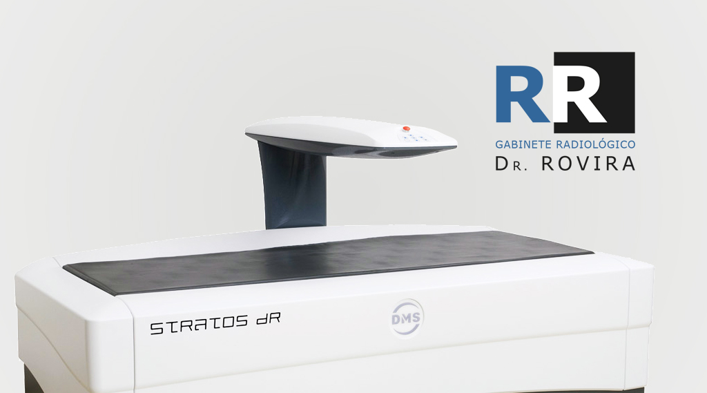
On the central DXA exam, which measures bone density in the hip and spine, the patient lies on a padded table. An x-ray generator is located below the patient and an imaging device, or detector, is positioned above.
To evaluate the spine, the patient’s legs are supported by a padded box to flatten the pelvis and the lower (lumbar) part of the spine. To evaluate the hip, the patient’s foot is placed on a clamp that rotates the hip inward. In both cases, the detector slowly passes through the area, generating images on a computer monitor.
You should remain still and you may be asked to hold your breath for a few seconds while taking the X-ray image to reduce the chance of it being blurred. The technician will head behind a wall or into the next room to activate the X-ray machine.
Peripheral exams are simpler. The finger, hand, forearm, or foot is placed in a small device that takes a bone density reading in a few minutes.
The DXA bone density test is usually done in 10 to 15 minutes, depending on the equipment used and the body parts examined.
Bone density tests are quick and painless. On the day of the exam you can eat normally. You should not take calcium supplements for at least 24 hours before the exam.
You should wear comfortable, loose clothing, avoiding clothes that have metal clasps, belts, or buttons. Objects such as keys or wallets that may be in the area to be examined should be removed. You may be asked to remove all or part of your clothing and to wear a gown during the exam. You may also be asked to remove jewelry, dentures, glasses, and any metal objects or clothing that may interfere with X-ray images.
Tell your doctor if you have recently had a barium examination or have had contrast dye injected for a computed tomography (CT) scan or radioisotopy. You may have to wait 10 to 14 days before taking the DXA test.
Women should always inform their doctor and X-ray technician if there is a possibility of pregnancy.
Today, the Rovira Cabinet has new professionals, new infrastructure and, as always, the best care and service in Diagnostic by image.
© 2020, Dr. Rovira Clinic. All rights reserved.